Anterior Shoulder Tendon Anatomy / Subscapularis Muscle Isolated Shoulder Anatomy Anterior View On Black Background Stock Photo .... All of them are supplied by the respective branches of the brachial plexus. Anterior fibers flex & medially rotate arm; Deltoid tuberosity of the humerus. Your rotator cuff consists of the muscles and tendons in your shoulder. The bursa can become inflamed and swell with more fluid causing pain.
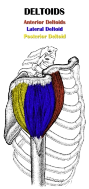
Its main function is shoulder flexion, which is characterized by raising your upper arms up to the front and overhead. The shoulder joint (glenohumeral joint) is a ball and socket joint between the scapula and the humerus. All of them are supplied by the respective branches of the brachial plexus. Posterior fibers extend & laterally rotate arm. Shoulder injuries like fractures bone breakage injured tendons or muscles are the typical causes of anterior shoulder pain.
Shoulder muscles : Anatomy and functions | Kenhub from thumbor.kenhub.com Below are some anatomic terms doctors use to describe location (as applied to the shoulder): The rotator cuff tendons can be irritated or damaged. The bursa can become inflamed and swell with more fluid causing pain. The rotator cuff is a collection of muscles and tendons that surround the shoulder, giving it support and allowing a wide range of motion. These contribute, together with the subscapularis, supraspinatus, infraspinatus and teres minor muscles, to the stability of the joint. The scapula, clavicle and humerus are the bones of the shoulder. Tendonitis of your shoulder is an inflammation of your rotator cuff or biceps tendon. It works to allow a lot of range of motion in forward flexion (arms in front.
A tendon is a structure that connects muscle to bone, and the biceps are connected by tendons at both the elbow and shoulder joints.
Anterior shoulder tendon anatomy : The shoulder region includes the glenohumeral joint, the. Three bones come together at the shoulder joint. The anterior deltoid is located on the front of your shoulder. It works to allow a lot of range of motion in forward flexion (arms in front. The shoulder isn't just one bone, it's actually made up of three different bones and various tendons, ligaments, and muscles.the three bones located in the shoulder are the humerus, the scapula, and the clavicle. Pectoralis major is a large muscle located on the anterior chest wall that has several shoulder joint related functions. In this condition the rotator cuff is unable to support the glenohumeral joint thereby causing pain in the biceps and the shoulder. Shoulder tendon anatomy (page 1). Also in its upper part, the anterior portion of the subacromial subdeltoid bursa can be seen deep to the deltoid muscle and anterior to the biceps sheath. It is an injury to the glenohumeral joint (ghj) where the humerus is displaced from its normal position in the center of the glenoid fossa and the joint surfaces no longer touch each other. The anatomy of the lhbt and its corresponding structures has been extremely well researched. The bicipital groove of the humerus.
Beyond this, there is also a shoulder joint arrayed in a ball and socket formation, a rotator cuff, and various muscles like the deltoid muscle and the teres major muscle. The bursa is a small sac of fluid that cushions and. The shoulder joint (glenohumeral joint) is a ball and socket joint between the scapula and the humerus. The glenohumeral joint is the main joint and is more like a golf ball sitting on a tee. It works to allow a lot of range of motion in forward flexion (arms in front.
Shoulder Anatomy | All About the Shoulder Muscles from www.kingofthegym.com The shoulder region includes the glenohumeral joint, the. The bursa is a small sac of fluid that cushions and. Deltoid tuberosity of the humerus. Shoulder anatomy for ultrasound evaluation. A tendon is a structure that connects muscle to bone, and the biceps are connected by tendons at both the elbow and shoulder joints. The anatomy of the lhbt and its corresponding structures has been extremely well researched. The long head of biceps (lhb) is a very important tendon that travels through the shoulder joint (glenohumeral joint).the biceps tendon begins at the top of the shoulder socket (the glenoid) and then passes across the front of the shoulder to connect to the biceps muscle. Your rotator cuff consists of the muscles and tendons in your shoulder.
The rotator cuff tendons can be irritated or damaged.
Posterior fibers extend & laterally rotate arm. Beyond this, there is also a shoulder joint arrayed in a ball and socket formation, a rotator cuff, and various muscles like the deltoid muscle and the teres major muscle. 11 photos of the shoulder muscles tendons anatomy. These contribute, together with the subscapularis, supraspinatus, infraspinatus and teres minor muscles, to the stability of the joint. Below are some anatomic terms doctors use to describe location (as applied to the shoulder): The long head of biceps (lhb) is a very important tendon that travels through the shoulder joint (glenohumeral joint).the biceps tendon begins at the top of the shoulder socket (the glenoid) and then passes across the front of the shoulder to connect to the biceps muscle. Biceps tendons the biceps muscle has two tendons at the shoulder, called the long head and short head. Anterior shoulder muscles, also called the pectoral muscles, attach the upper extremity to the clavicle and the thoracic cage. They connect your upper arm bone to your shoulder blade. It is an injury to the glenohumeral joint (ghj) where the humerus is displaced from its normal position in the center of the glenoid fossa and the joint surfaces no longer touch each other. The term anterior shoulder instability refers to a shoulder in which soft tissue or bony insult allows the humeral head to sublux or dislocate from the glenoid fossa. The anterior limb of the circumflex humeral artery is frequently visible around the tendon. Anterior shoulder tendon anatomy :
Axillary nerve (c5,6) from posterior cord of brachial plexus. Posterior fibers extend & laterally rotate arm. This muscle works in combination with pectoralis minor which lies underneath it. On the anterior side of the shoulder, the coracobrachialis, serratus anterior, pectoralis major, and pectoralis minor muscles work as a group to flex and adduct the scapula and humerus anteriorly toward the sternum. Shoulder tendons chart ~ labeled anatomy chart of shoulder ligaments on white background stocktrek images.
Muscles of the Shoulder and Arm Anterior View - Medical Stock Images Company from cdn.shopify.com The bicipital groove of the humerus. Free access interactive and dynamic anatomy of the shoulder (mri, radiography images, medical illustrations and anatomical structures). In this condition the rotator cuff is unable to support the glenohumeral joint thereby causing pain in the biceps and the shoulder. Biceps tendons the biceps muscle has two tendons at the shoulder, called the long head and short head. Injuries resulting from dysfunction are common and potentially debilitating. Anterior shoulder muscles, also called the pectoral muscles, attach the upper extremity to the clavicle and the thoracic cage. Axillary nerve (c5,6) from posterior cord of brachial plexus. The latissimus dorsi and teres major on the posterior side extend and adduct the arm towards the vertebrae of the back.
Beyond this, there is also a shoulder joint arrayed in a ball and socket formation, a rotator cuff, and various muscles like the deltoid muscle and the teres major muscle.
Anterior shoulder muscles, also called the pectoral muscles, attach the upper extremity to the clavicle and the thoracic cage. It is an injury to the glenohumeral joint (ghj) where the humerus is displaced from its normal position in the center of the glenoid fossa and the joint surfaces no longer touch each other. Your rotator cuff consists of the muscles and tendons in your shoulder. The shoulder is extremely mobile and made up of several joints that work together. The shoulder region includes the glenohumeral joint, the. Deltoid tuberosity of the humerus. The head of the humerus usually tears the inferior part of the joint capsule because this region is the least protected part of the capsule. They connect your upper arm bone to your shoulder blade. The shoulder joint is composed of the glenoid (the shallow shoulder socket) and the head of the upper arm bone known as the humerus (the ball). The glenohumeral joint is the main joint and is more like a golf ball sitting on a tee. Beyond this, there is also a shoulder joint arrayed in a ball and socket formation, a rotator cuff, and various muscles like the deltoid muscle and the teres major muscle. The acromion can rub against (or impinge on) the tendon and the bursa, causing irritation and pain. The anterior limb of the circumflex humeral artery is frequently visible around the tendon.
Its main function is shoulder flexion, which is characterized by raising your upper arms up to the front and overhead shoulder tendon anatomy. Also in its upper part, the anterior portion of the subacromial subdeltoid bursa can be seen deep to the deltoid muscle and anterior to the biceps sheath.

0 Comments:
Posting Komentar