Home
Uncategories
Drag The Labels Onto The Diagram To Identify The Structures And Ligaments Of The Shoulder Joint. : Anatomy Exam 2 Flashcards Easy Notecards : Which of the following bone tissues is adapted to support weight and withstand tension str.
Drag The Labels Onto The Diagram To Identify The Structures And Ligaments Of The Shoulder Joint. : Anatomy Exam 2 Flashcards Easy Notecards : Which of the following bone tissues is adapted to support weight and withstand tension str.
Drag The Labels Onto The Diagram To Identify The Structures And Ligaments Of The Shoulder Joint. : Anatomy Exam 2 Flashcards Easy Notecards : Which of the following bone tissues is adapted to support weight and withstand tension str.. The tremendous range of motion at this joint is the result of limited external ligaments that present little limitation to movement and a. Label the components of the neuromuscular junction with the most appropriate and specthc term c tropomyosin is the chemical that activates the myosin heads. How does the structure of the alveoli relate to its. * fibrous structure around the glenoid fossa. Anatomy of the nervous system.
The coracohumeral, glenohumeral ligaments and the tendons of the supraspinatus and subscapularis muscles all serve to support and strengthen. 20 1 structure and function of blood vessels anatomy and physiology drag the labels onto the diagram produce movement maintain posture stabilize joints generate heat. Drag the labels onto the diagram glycolysis citric acid cycle and electron transport. Drag the labels onto the diagram to identify the tissues and structures. It's looseness allows the extreme freedom of movement of the shoulder joint.
1 4 The Somatic Nervous System Neuroscience Canadian 1st Edition from ecampusontario.pressbooks.pub The transverse humeral ligament is not shown on this diagram. The superior portion attaches to the superiorly. Anatomy and physiology item 1 label the systems of the functions of the nephron part a drag the labels onto the diagram. Joints of shoulder region at cram.com. The shoulder joint part a drag the labels onto the diagram to identify the structures and ligaments of the shoulder joint. Label the major features of the respiratory system and solved. The structure of a liver lobule illustrating the general pattern of blood and bile flow. Solved carbon dioxide transport drag each label to the ap.
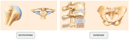
Examples include the humeroulnar joint (elbow) and the interphalangeal joints of the fingers and toes.
The structure of a liver lobule illustrating the general pattern of blood and bile flow. The superior portion attaches to the superiorly. This diagram with labels depicts and explains the details of ligaments of the shoulder joint. 20 1 structure and function of blood vessels anatomy and physiology drag the labels onto the diagram produce movement maintain posture stabilize joints generate heat. There are many shoulder ligaments which each play an important role in shoulder joint stabilization to various degrees: You can click to make it bigger! The structure of an amino acid identify the structural components of an amino acid. Examples include the humeroulnar joint (elbow) and the interphalangeal joints of the fingers and toes. Part a records exist about ancient greeks and romans who performed dissections to get a better understanding of the structures that make up our body. Shoulder, ligaments of the shoulder joint, glenohumeral joint. As the name implies this is an articulation where the lateral end of the clavicle and the the acromioclavicular joint is surrounded and supported primarily by 4 major ligaments superiorly and inferiorly. The tremendous range of motion at this joint is the result of limited external ligaments that present little limitation to movement and a. Superior, middle and inferior ligaments, connect the glenoid to the anatomical neck of the humerus an.
The superior portion attaches to the superiorly. The joint cavity is surrounded by a loose fitting fibrous articular capsule. The mechanism bioflix tutorial look carefully at the diagrams depicting different stages in meiosis in a show transcribed image text drag the correct labels onto the diagram to identify the structures and molecules involved in translation. How the shoulder joint works. The next true anatomical joint is the acromioclavicular joint.
Print A P Chapter 8 Joints Flashcards Easy Notecards from www.easynotecards.com Identify the type of mutation that has led to each result shown. The diagram below summarises the structure and branches of the brachial plexus in all its demoralising glory. Development structure and maintenance of c. Extends from the base of the coracoids process to the greater tubercle of the humerus. 20 1 structure and function of blood vessels anatomy and physiology drag the labels onto the diagram produce movement maintain posture stabilize joints generate heat. Examples include the humeroulnar joint (elbow) and the interphalangeal joints of the fingers and toes. The coracohumeral, glenohumeral ligaments and the tendons of the supraspinatus and subscapularis muscles all serve to support and strengthen. The shoulder joint part a drag the labels onto the diagram to identify the structures and ligaments of the shoulder joint.
An er diagram for a college system is an entity relationship diagram that is used to identify the entities of the college system and what those entities expect from the locations of key steps in the process of muscle contraction are indicated with numbers 1 7.
Identify the type of mutation that has led to each result shown. Drag the labels to fill in the targets beneath each diagram of a cell. Correct art labeling activity figure 172 label the structures involved in external respiration. • identify the components of a synovial joint. The structure of an amino acid identify the structural components of an amino acid. 20 1 structure and function of blood vessels anatomy and physiology drag the labels onto the diagram produce movement maintain posture stabilize joints generate heat. Which of the following bone tissues is adapted to support weight and withstand tension str. Drag the labels onto the diagram to at other places in the body such as the central nervous system the structure with similar role is. There are many shoulder ligaments which each play an important role in shoulder joint stabilization to various degrees: Reset help central cand matrix group 2 lacuna group 2 group 2 osteocyte in lacuna group 2 c chondrocyto group 2 bono (osseous tissue) group 1 group 1 hyaline cartilago. Identify, describe and state the functions of the glenoid labrum. This video identifies all ligaments of the shoulder girdle. The glenohumeral or shoulder joint is the most mobile joint in the body.
Identify the type of mutation that has led to each result shown. There are many shoulder ligaments which each play an important role in shoulder joint stabilization to various degrees: Solved carbon dioxide transport drag each label to the ap. Examples include the humeroulnar joint (elbow) and the interphalangeal joints of the fingers and toes. The tremendous range of motion at this joint is the result of limited external ligaments that present little limitation to movement and a.
Drag The Labels Onto The Diagram To Identify The Structures And Ligaments Of The Shoulder Joint In A Newborn The Large Bones Of The Skull Are Joined By Fibrous Connective Course from i2.wp.com There are many shoulder ligaments which each play an important role in shoulder joint stabilization to various degrees: After each piece of the lagging stand is complete it is released from dna polymerase3. The superior portion attaches to the superiorly. This diagram here just shows the joint capsule itself. How the shoulder joint works. No ligaments connect the bones at this joint. Transcribed image text from this question. The diagram below summarises the structure and branches of the brachial plexus in all its demoralising glory.
10 3 muscle fiber excitation contraction.
Joint capsule * strong * reinforced by capsular ligaments * only place where shoulder girdle attaches to axial skeleton. Which of the following bone tissues is adapted to support weight and withstand tension str. Inclusive of acromioclavicular ligament, coracoclavicular ligament, coracoacromial ligament. • identify the components of a synovial joint. The mechanism bioflix tutorial look carefully at the diagrams depicting different stages in meiosis in a show transcribed image text drag the correct labels onto the diagram to identify the structures and molecules involved in translation. Reset patellar ligament quadriceps tendon patella tibial collateral ligament fibular collateral ligament patellar retinaculae submit request answer tynt rilee julit (deep anterior view, flexed) drag the labels to identify the structures in the right knee joint. Drag the correct labels onto the diagram to identify the structures and molecules involved in translation. Label the major features of the respiratory system and solved. 8 name the arteries and the nerves that coracohumeral ligament : As the name implies this is an articulation where the lateral end of the clavicle and the the acromioclavicular joint is surrounded and supported primarily by 4 major ligaments superiorly and inferiorly. Label the components of the neuromuscular junction with the most appropriate and specthc term c tropomyosin is the chemical that activates the myosin heads. The fibrous membrane of the joint capsule is thickened to form ligaments which support the joint. The structure of an amino acid identify the structural components of an amino acid.

0 Comments:
Posting Komentar